Our knowledge about concepts such as body scheme, personal space, and near space perception has grown tremendously over the recent years. This has largely been due to a broad array of neurophysiological, neurokinesiological, and neuropsychological research evidence. Bringing these concepts down to the practical level of function and intervention for the child with SPD can be daunting.
However, given advances in fmri and DTI neuroimaging practices, better understanding of the integrated brain pathways that support functions of spatial perception have emerged. Although the key players of functional brain pathways in the development of body awareness have not changed, awareness of the changing roles of these key players has certainly been expanded. While the researchers still seem to agree that the vestibular, visual, and tactile-proprioceptive systems remain major players, their involvement in the coding of spatial orientation seems to have shifted.
One thought-provoking point brought into the arena is that both the visual and the vestibular systems have been shown to play a role in increasing somatosensory gain sensitivity. Clinically, this means that when vision and/or vestibular input are brought into the equation, children with SPD who have deficits in body awareness should perform better when these types of additional input are included.
Another thought-provoking point brought into the arena is that researchers have found that children with Developmental Coordination Disorder may require larger sensory discrepancies in order to register error signals. Clinically, this means that rather than initially focusing upon the ability to discriminate among small differences, children with SPD should be better able to adapt and perform better when larger somatosensory differences are introduced.
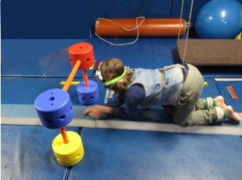
 We used these lines of clinical reasoning when we set up this particular motor maze. We wanted the youngster to be able to balance, bringing all four extremities into a point of self representation of movement on the beam. Yes, he had to narrow his base of support, and pre-plan for moves of each limb prior to movement in order to stay erect upon the beam.
 In addition, we wanted him to become more aware of “near space”, adapting his presence to environmental cues and obstacles of the environment. We were particularly interested to see whether or not these types of visual and vestibular influences would act as “gain regulators” as reported by researchers to address greater efficiency in his somatosensory processing skills
 We took care to select oversized equipment as we planned to observe whether or not he would be better able to adapt to somatosensory cues when given larger fields of discrepancy as he navigated the obstacles. We used this activity for two weeks over four treatment sessions, while varying the follow-up table top activity each time.
Although our project was not set up to provide formal outcome measures, we were incredibly pleased to see much improved postural control and motor planning emerge. This was spontaneously shown a week later when this youngster was able to mount and skillfully use equipment that heretofore had been hazardous for him.
References:
Jody C. Culham, et. al: The Role of parietal cortex in visuomotor control: What have we learned from neuroimaging?
Elisa Ferre, et. al: Multisensory Interactions between Vestibular, Visual and Somatosensory Signals
http://journals.plos.org/plosone/article?id=10.1371/journal.pone.0124573
Mitsuru Kashiwagi: Parietal dysfunction in developmental coordination disorder: a functional MRI study.

Comments are closed.